How the immune system senses infection and danger.
Pattern recognition receptors (PRRs) are germline-encoded sensors of the innate immune system. They recognize conserved molecular patterns from microbes (pathogen-associated molecular patterns, PAMPs) and signals released by damaged or stressed cells (damage-associated molecular patterns, DAMPs), helping to initiate innate defenses and shape adaptive immunity.
Induce inflammatory cytokines and type I interferons.
C-type lectin receptors(CLRs)
Cell surface
Recognize carbohydrate structures (e.g., β-glucans from fungi).
Important for antifungal immunity and also sense self-derived ligands.
Activate NF-κB and other pathways.
NOD-like receptors(NLRs)
Cytosol
Sense bacterial components (e.g., peptidoglycan) and danger signals.
Some NLRs (e.g., NLRP3) form inflammasomes.
Activate caspase-1 and promote IL-1β and IL-18 production.
RIG-I-like receptors(RLRs)
Cytosol
Detect viral RNA in the cytosol.
Activate IRF3/IRF7 and NF-κB pathways.
Induce type I interferons and antiviral responses.
AIM2-like receptors(ALRs)
Cytosol
AIM2 detects cytosolic DNA.
Forms an inflammasome with caspase-1.
Induces IL-1β and IL-18 and can trigger pyroptosis.
cGAS–STING pathway
Cytosol
cGAS senses cytosolic DNA and produces cGAMP.
STING activates IRF3 and NF-κB pathways.
Induces type I interferons and inflammatory cytokines.
From recognition to immune response
PRR signaling triggers a coordinated sequence of events that link innate and adaptive immunity.
1Detection of PAMPs or DAMPs
PRRs recognize conserved microbial or danger signals at the cell surface, in endosomes or in the cytosol.
2Receptor activation and signaling
Adaptor proteins
Recruitment of adaptor proteins (e.g., MyD88, TRIF, STING) and activation cascades.
3Activation of transcription factors
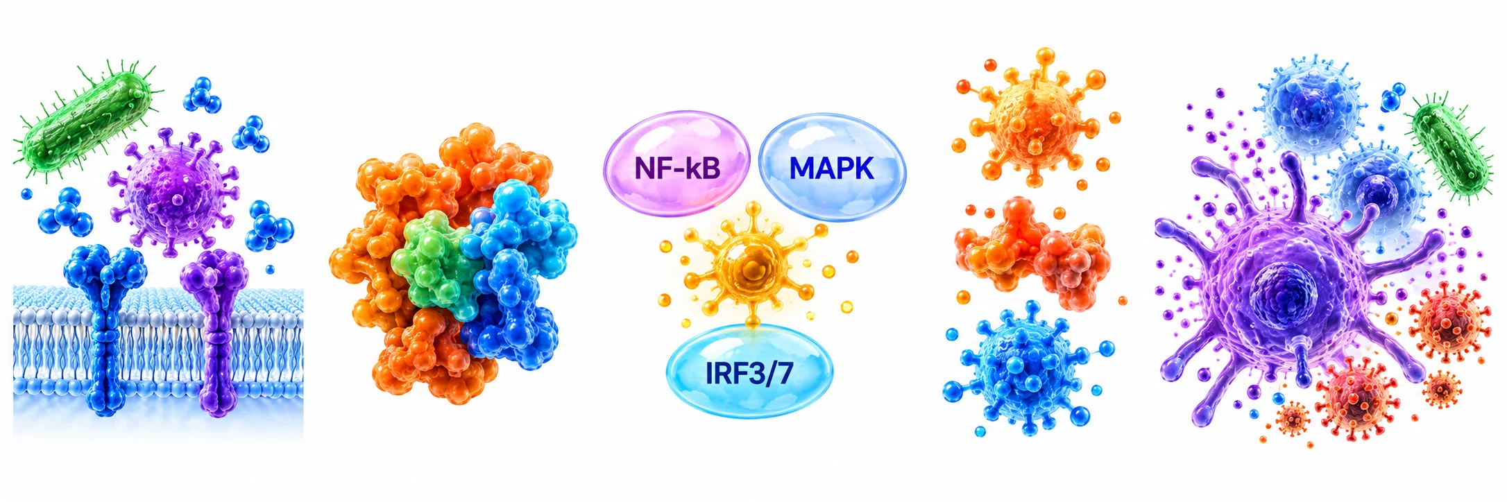
Activation of NF-κB, MAPK and/or IRF3/7 leads to changes in gene expression.
4Production of immune mediators
Cytokines (e.g., TNF-α, IL-6) Chemokines (e.g., CCL2, CXCL10) Type I interferons (IFN-α/β)
5Immune response and link to adaptive immunity
Recruitment and activation of immune cells and enhanced antigen presentation help initiate adaptive immunity.
Where PRRs are located in the cell
Why PRRs matter
PRRs provide essential protection and help maintain overall health.
Rapid detection
Recognize microbial and danger signals before infection is established.
Fight infections
Activate antimicrobial responses and antiviral defenses.
Initiate inflammation
Drive production of cytokines and chemokines to recruit immune cells.
Bridge to adaptive immunity
Help activate dendritic cells and enhance T and B cell responses.
Sense tissue damage
Detect danger signals from stressed or injured cells and help with tissue repair.
When PRR signaling goes wrong
Imbalanced PRR activation can contribute to disease.
Excessive or persistent activation
Can contribute to chronic inflammation and immune-mediated diseases (e.g., autoimmune disease, cardiovascular disease, metabolic disorders).
May be driven by persistent infection, sterile inflammation or dysregulated self-sensing.
Insufficient or impaired signaling
Can lead to increased susceptibility to infections (e.g., severe viral or bacterial infections).
Genetic deficiencies in PRR pathways can cause primary immunodeficiencies.
PRRs and aging
Aging can alter innate immune sensing and PRR signaling, contributing to a state of low-grade inflammation (inflammaging) and changes in responses to infections and vaccines.

 Subscribe