Skip to content

Major Histocompatibility Complex

How cells show antigens to T cells

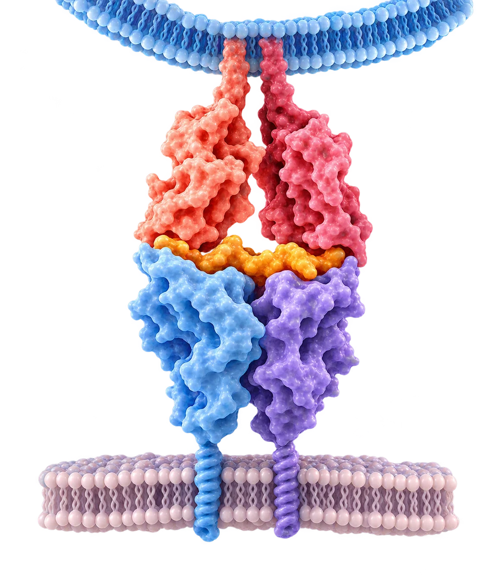
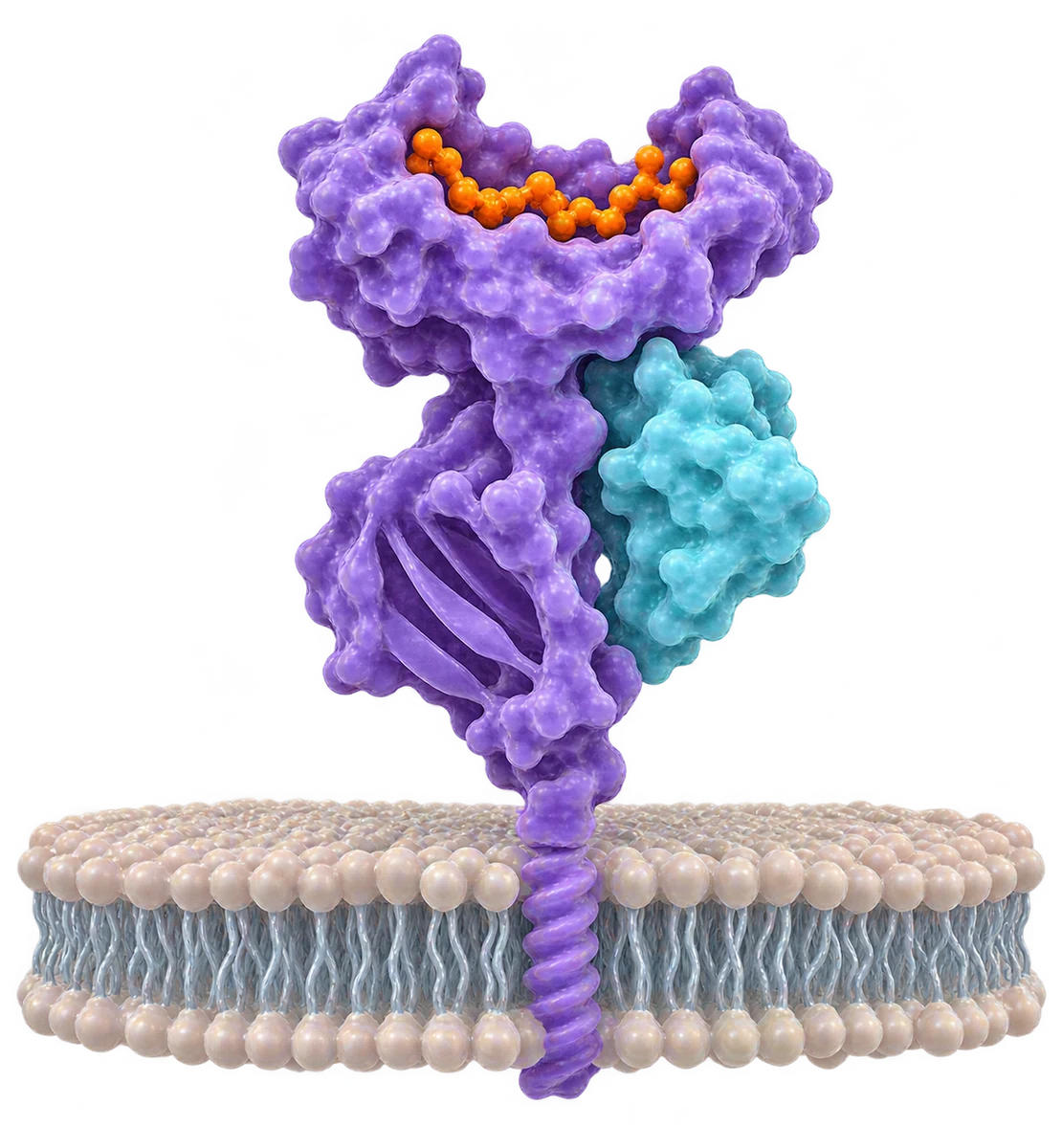
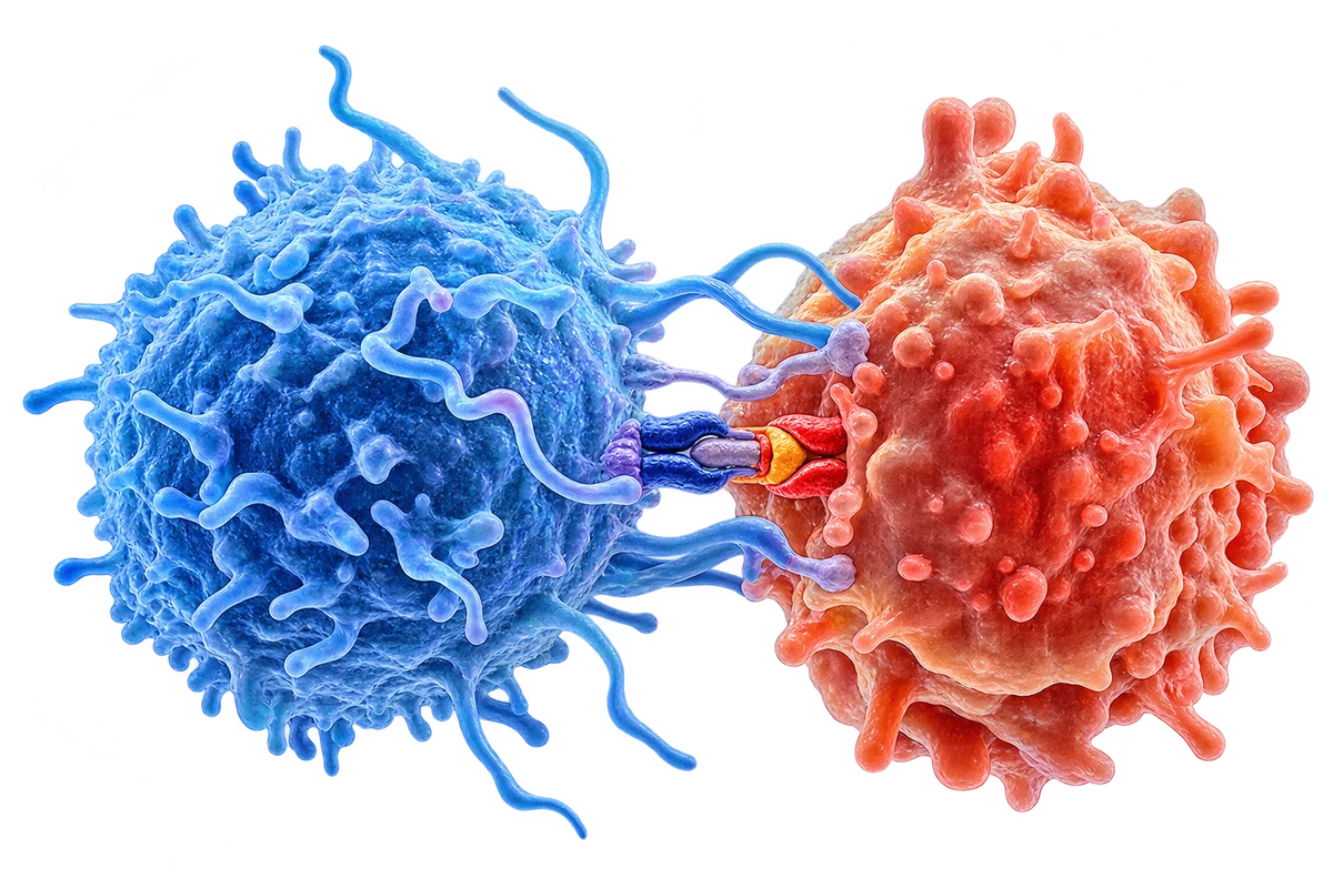
Major histocompatibility complex (MHC) molecules display small protein fragments called peptides. T cells recognize these peptide–MHC complexes through their T-cell receptors. In humans, MHC molecules are called HLA—human leukocyte antigens.

Explore MHC and antigen presentation →

Two pathways, different T cells

MHC Class I

Displays peptides from inside the cell
to CD8+ T cells

  • Expressed on most nucleated cells.
  • Mainly presents peptides derived from intracellular proteins.
  • Recognized by CD8+ T cells.
  • Human classical genes: HLA-A, HLA-B and HLA-C.
MHC I presenting a peptide to the T-cell receptor of a CD8+ T cells lymphocyte
Heavy chainβ₂-microglobulinPeptideT-cell receptor

MHC Class II

Displays peptides from proteins taken up by the cell to CD4+ T cells

  • Mainly expressed by dendritic cells, macrophages and B cells.
  • Mainly presents peptides from internalized proteins.
  • Recognized by CD4+ T cells.
  • Human classical molecules: HLA-DP, HLA-DQ and HLA-DR.
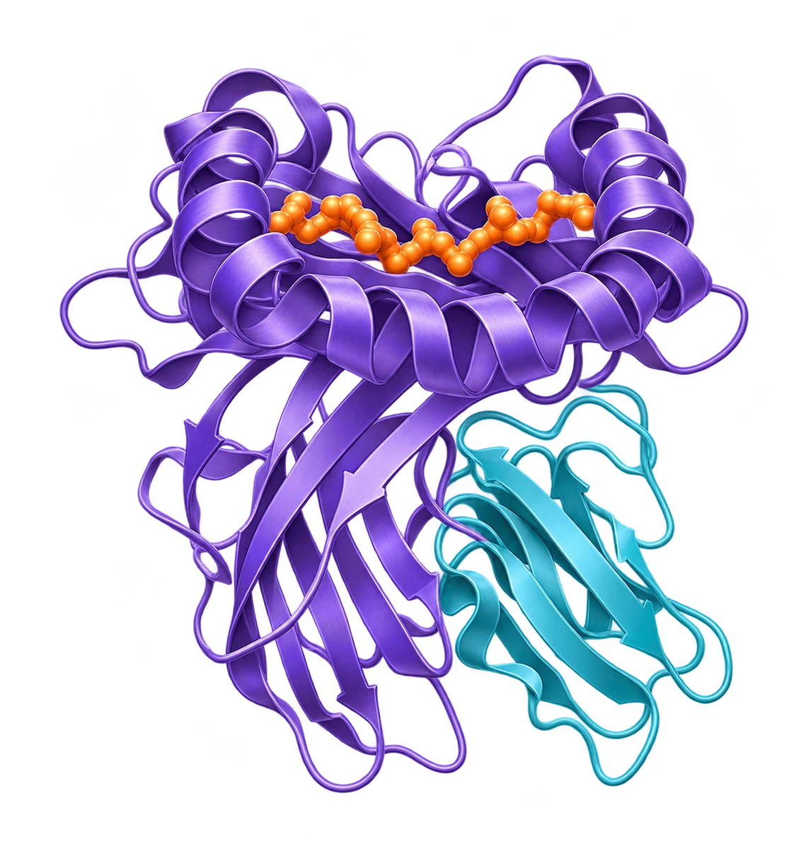
MHC II presenting a peptide to the T-cell receptor of a CD4+ T cells lymphocyte
α chainβ chainPeptideT-cell receptor

From protein to presentation

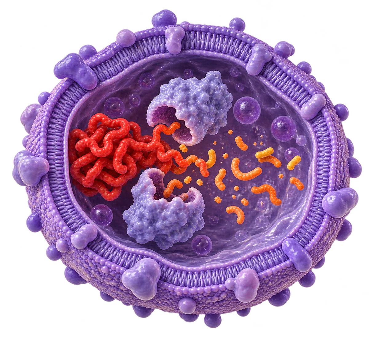
The main routes of peptide processing and display.

MHC Class I

(endogenous pathway)

Intracellular protein

Intracellular protein

Proteins made within the cell provide substrates for processing.

Proteasome

Proteasome

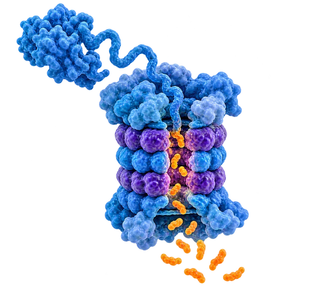
Proteasomes generate peptide fragments in the cytosol.

TAP transport into ER

TAP transport into ER

TAP transports peptides into the endoplasmic reticulum.

Peptide loading in ER

Peptide loading in ER

Suitable peptides bind newly assembled MHC I molecules.

Cell surface

Cell surface

Peptide–MHC I complexes travel to the surface for CD8+ T-cell recognition.

MHC Class II

(exogenous pathway)

Antigen uptake

Antigen uptake

Antigen-presenting cells internalize extracellular material.

Endosome / lysosome

Endosome / lysosome

Proteases break down proteins in acidified compartments.

Peptide loading

Peptide loading

HLA-DM supports peptide exchange on MHC II after invariant-chain processing.

Cell surface

Cell surface

Peptide–MHC II complexes reach the surface for CD4+ T-cell recognition.

An important exception

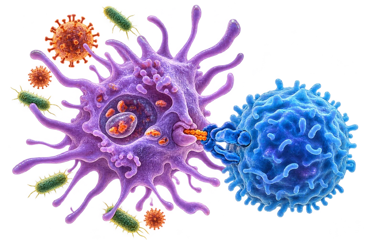
Dendritic-cell cross-presentation to a CD8 T cell

Dendritic cells can present peptides from captured extracellular antigens on MHC I through cross-presentation. This helps prime CD8+ T-cell responses. MHC II can also receive some intracellular proteins through additional pathways.

Recognition is only the first step

T-cell receptor recognition, co-stimulation and cytokine signals

Activation of naive T cells usually also requires co-stimulatory signals and an appropriate cytokine environment. Recognition alone does not guarantee an effective immune response.

MHC & Aging

Aging can alter antigen uptake, processing, dendritic-cell signaling and T-cell priming. Findings vary with cell type, tissue and experimental conditions.

More than MHC levels

MHC expression alone does not reveal how effectively a cell presents antigen. Co-stimulation, cytokines and T-cell function also matter.

Age-related changes in antigen presentation are not a uniform decline in MHC expression.

Learn about dendritic cells →
Older adult in a mountain setting

Why it matters

Infection defense

Infection defense

Peptide–MHC recognition helps T cells identify infected cells and coordinate pathogen-specific responses.

Learn more →
Vaccine responses

Vaccine responses

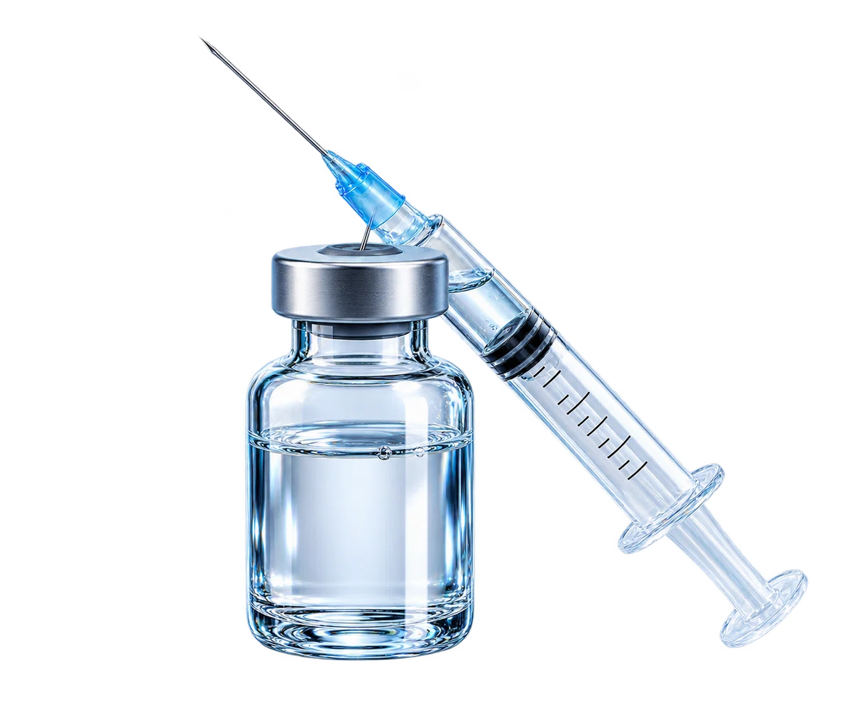
Many vaccines support protective immunity by generating antigen presentation and subsequent T- and B-cell responses.

Learn more →
Immune recognition

Immune recognition

T cells survey peptide–MHC complexes. Responses depend on antigen recognition together with tolerance and regulatory signals.

Learn more →

Peptide–MHC recognition is fundamental to adaptive immunity.

Selected sources and references

Artwork illustrates the main mechanisms schematically; molecular sizes, colors and distances are not literal.