1. Bind IgE
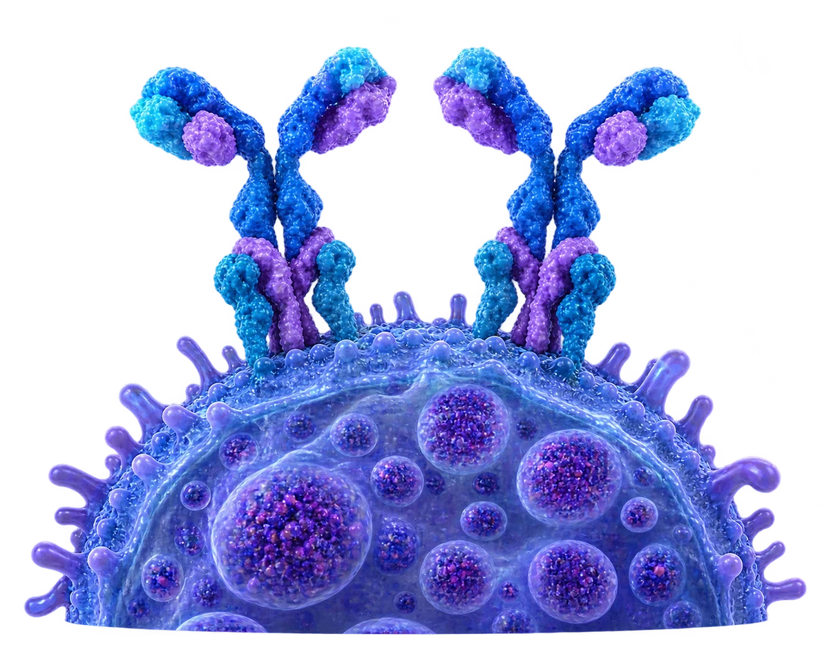
IgE binds high-affinity FcεRI receptors on the basophil surface.
 Subscribe
IMMUNE CELLS
Basophils are rare blood granulocytes with dense granules and high-affinity IgE receptors. They release histamine, lipid mediators and cytokines that influence allergic and type 2 immune responses. Their functions overlap with mast cells, but they have a distinct developmental pathway and tissue behavior.
Back to all innate immune cells →Basophils develop in bone marrow and normally make up less than 1% of circulating white blood cells. Large blue-purple granules partly obscure their lobed nucleus. Unlike mast cells, they mature primarily in marrow and circulate before being recruited to tissues.
Basophils express high-affinity FcεRI receptors that bind IgE. Appropriate stimulation can release histamine and lipid mediators and induce cytokines such as IL-4 and IL-13. These signals influence other cells during allergic inflammation and some host-defense responses.
IgE-dependent activation is one pathway among several receptor and cytokine inputs.
IgE binds high-affinity FcεRI receptors on the basophil surface.

Allergen cross-linking and other signals can initiate an activation response.
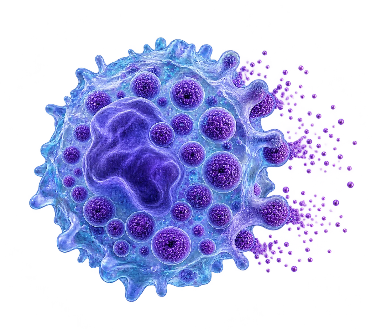
Stored histamine and newly generated lipid mediators affect local tissues.

IL-4 and IL-13 help shape responses in other immune and tissue cells.
Specialized cellular functions contribute to coordinated immunity.
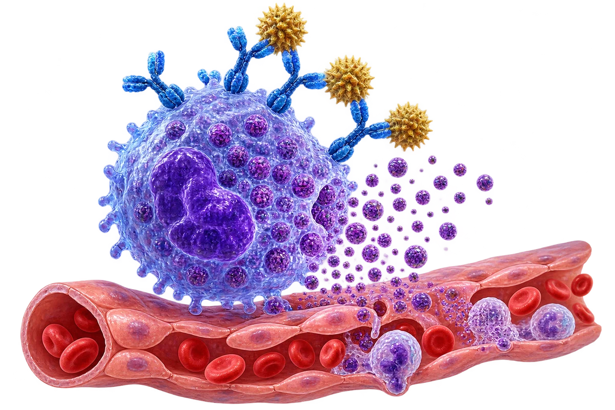
IgE-dependent and other pathways can drive mediator release in allergic responses.
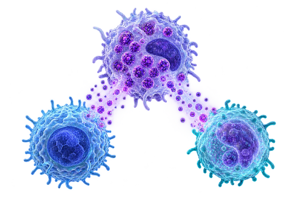
IL-4 and IL-13 influence helper-cell and tissue responses.
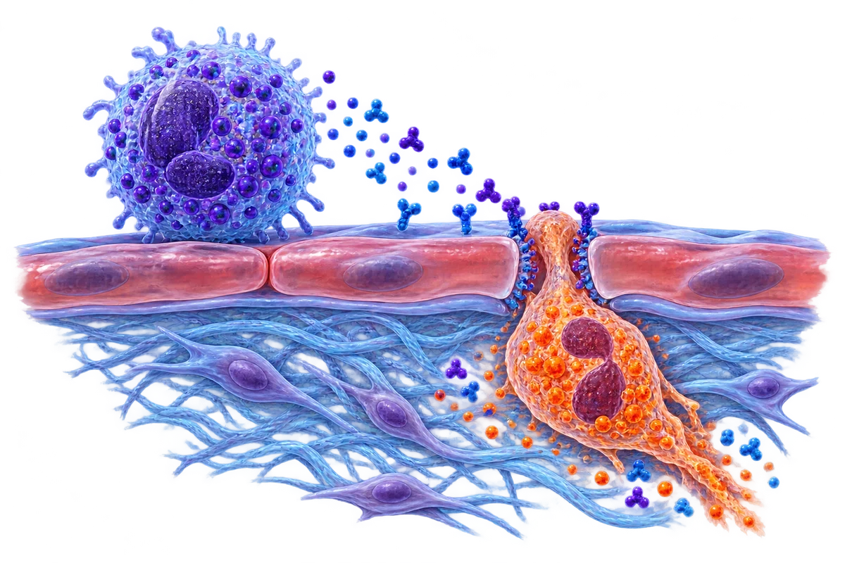
Mediators affect vascular and tissue signals that guide other leukocytes.
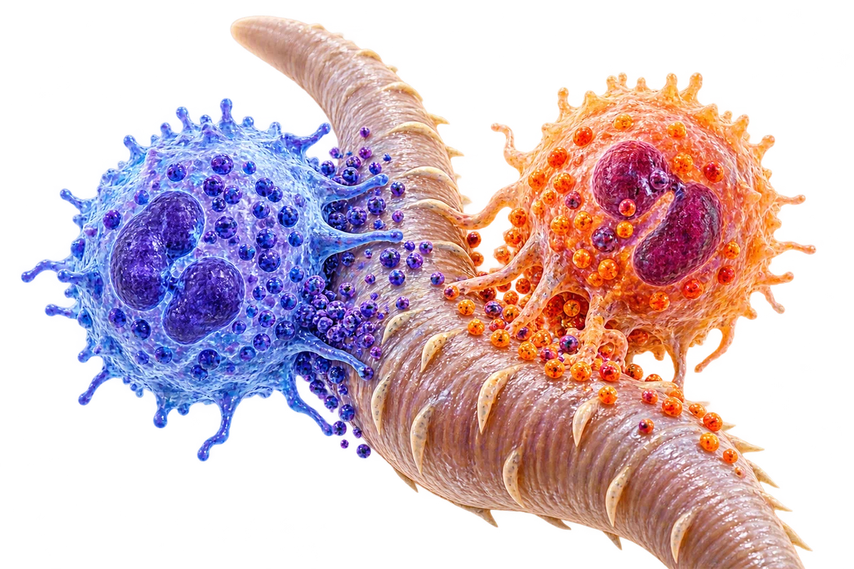
Basophils contribute to some parasite-related responses in a context-dependent way.

Direct evidence on human basophil aging remains limited compared with other immune cells.
Artwork illustrates the main mechanisms schematically; molecular sizes, colors and distances are not literal.